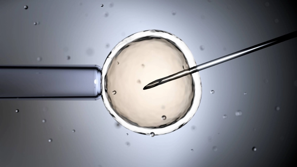
A team led by a professor at Osaka University's Bioinformatics Center has replicated the mouse embryo implantation process in laboratory equipment with a high success rate.

Masahito Ikawa and his team on Wednesday said that they had developed a method of culturing fragments of uterine tissue from mice in laboratory containers to achieve embryo implantation.

The team also discovered protein interactions that help drive embryo implantation.

Its findings were published in the British scientific journal Nature Communications.

"We hope to develop a method to reduce implantation failure as much as possible in assisted reproductive technology for humans as well as a therapeutic drug" through the developed technique, Ikawa said.

Due to the lack of examination over the exact moment an implantation occurs within a human body, factors that determine the success or failure of implantation remain unclear.

For this reason, experiments have been conducted in containers to replicate such implantation. There are, however, ethical issues with using a large number of human in vitro embryos.

Researchers both in and outside of Japan are conducting experiments to produce model blastocysts, tiny cells that form soon after a fertilized egg starts dividing, mainly from induced pluripotent stem, or iPS, cells, and implant such fertilized eggs into the endometrium extracted from sick patients.

One of the advantages of experiments using mice is that it's easy to manipulate genes and administer drugs.

For their study, Ikawa and other members of his team built off a method of growing endometrial fragments extracted from female mice in containers while supplying oxygen. The team's technology to implant blastocysts had a success rate of some 95%.

Up until now, the specific function of COX-2, an enzyme in the uterine lining that promotes implantation, was unknown.

The team found through tests to inhibit enzyme activities with drugs and genetic analysis that COX-2 had activated a type of protein called AKT in cells outside the blastocysts.

Even when the function of COX-2 was impaired, progress was made with the implantation process once the artificially activated AKT was introduced into the blastocysts.